Canine dissection, guided by resources like Miller’s Anatomy, offers a detailed exploration of a dog’s anatomy, meticulously illustrating precise cutting techniques and key structures.
Historical Context of Canine Anatomy Study
Historically, the canine has served as a crucial model for anatomical study, bridging the gap between human and smaller animal physiology. Early anatomical investigations, predating modern imaging, relied heavily on dissection to understand mammalian structures. The dog’s anatomical similarities to humans made it invaluable for medical education and surgical training.
Landmark texts, such as those by Howard Evans and Alexander de Lahunta, have refined dissection guides over decades, building upon centuries of anatomical knowledge. These guides emphasize precise techniques and detailed anatomical terminology. The evolution of canine anatomy study reflects advancements in preservation methods – initially relying on simple drying, then progressing to formalin, phenol, and ethanol mixtures – enabling more thorough and lasting investigations.
Understanding this historical context is vital, as it demonstrates the enduring importance of hands-on anatomical exploration through dissection, solidifying the foundation for veterinary and medical advancements.
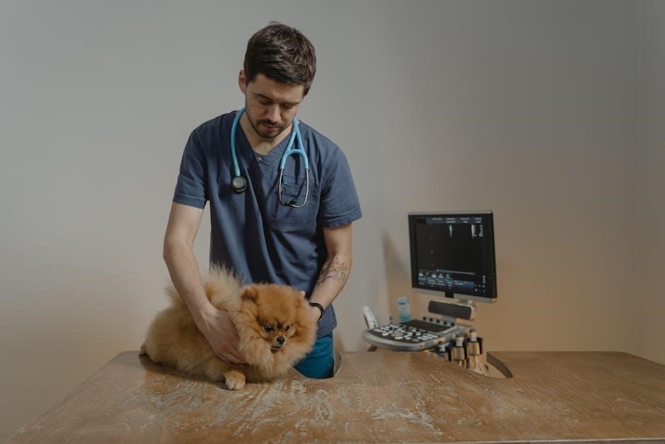
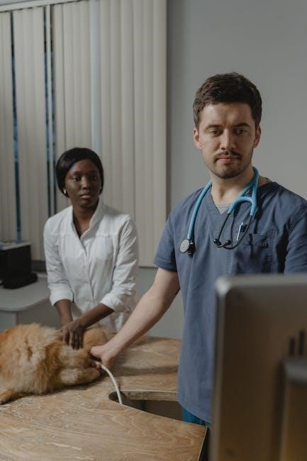
Importance of Dissection in Veterinary Education
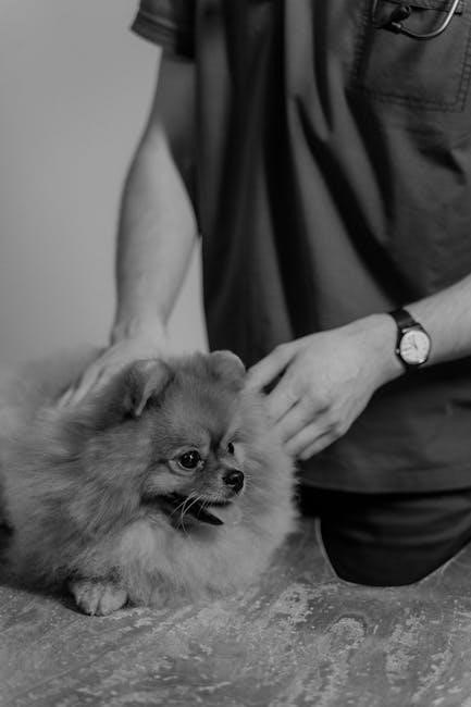
Dissection remains a cornerstone of veterinary education, providing an unparalleled three-dimensional understanding of canine anatomy that surpasses textbook illustrations and digital models. This hands-on experience fosters a crucial link between anatomical structures and their functional roles within the dog’s body.
Through careful dissection, students develop essential surgical skills, learning to identify and navigate delicate tissues, vessels, and nerves. The process reinforces anatomical terminology and spatial relationships, building a strong foundation for clinical diagnosis and treatment.
Furthermore, dissection cultivates a deeper appreciation for the complexity and interconnectedness of biological systems. It emphasizes the inseparable relationship between structure and function, a principle vital for effective veterinary practice. Utilizing guides like those based on Miller’s Anatomy ensures a systematic and comprehensive learning experience.
Preparation for Dissection
Prior to dissection, the dog undergoes humane euthanasia, typically via pentobarbital injection and exsanguination, followed by embalming with a formalin, phenol, and ethanol mixture.
Ethical Considerations and Humane Euthanasia
Ethical considerations are paramount when undertaking canine dissection, demanding respect for the animal and a commitment to minimizing suffering. The source dog must be obtained responsibly, often from facilities providing animals for scientific purposes, ensuring adherence to animal welfare regulations. Humane euthanasia is a critical first step, prioritizing a painless and stress-free passing.
The provided text details a common method: anesthesia induced by pentobarbital injection via the cephalic vein, followed by exsanguination through a cannula inserted into the common carotid artery. This sequence ensures complete blood removal before embalming, facilitating clearer anatomical visualization. Students must approach the dissection with reverence, recognizing the animal’s contribution to anatomical education and veterinary advancement. Acknowledging the ethical implications fosters a responsible and respectful learning environment.
Preservation Techniques: Formalin, Phenol, and Ethanol
Effective preservation is crucial for maintaining anatomical structures during canine dissection. A common embalming fluid, as detailed in the provided resources, utilizes a specific blend of chemicals to prevent decomposition and ensure long-term usability of the specimen. This solution typically consists of 5% formalin, 2% phenol, and 30% ethanol, carefully combined to achieve optimal preservation.
Formalin acts as a primary fixative, cross-linking proteins to stabilize tissues. Phenol contributes to tissue hardening and preservation of color, while ethanol serves as a carrier and aids in dehydration. This combination effectively halts bacterial growth and autolysis. Proper perfusion of the embalming fluid throughout the circulatory system is essential, ensuring thorough preservation of all organs and tissues for detailed anatomical study during dissection.
Necessary Dissection Tools and Equipment
Successful canine dissection requires a comprehensive set of specialized tools. Essential instruments include a sharp scalpel for precise incisions, various sizes of scissors – blunt and sharp – for careful tissue separation, and robust forceps, both hemostatic and dissecting, to grasp and manipulate structures. Retractors are vital for holding tissues apart, providing clear visibility during the dissection process.
Additionally, a bone saw or osteotome is necessary for sectioning bone, while probes aid in identifying and tracing vessels and nerves. A dissecting pan secures the specimen, and a good lighting source is paramount. Proper personal protective equipment, including gloves, lab coats, and eye protection, is non-negotiable for safety. Finally, labeling materials and a detailed anatomical guide, like Miller’s Anatomy, are indispensable for accurate identification and documentation.
Dissection of the Muscular System
Dog dissection involves carefully exposing and identifying muscles layer by layer, starting with superficial muscles of the limbs, then proceeding to deeper structures.
Superficial Muscle Dissection – Thoracic Limb
Begin the thoracic limb dissection by carefully making initial skin incisions, exposing the underlying fascia and superficial muscles. Systematically identify and separate muscles like the brachiocephalicus, trapezius, and omotransversarius, noting their origins, insertions, and functions.
Pay close attention to the cutaneous maximus and superficial pectoral muscles, carefully dissecting around neurovascular bundles to avoid damage. Observe the distinct fiber directions and muscle bellies, utilizing illustrations from anatomical guides for accurate identification.
As you progress, gently retract muscles to reveal deeper structures, documenting each step with precise labeling. Remember to correlate your observations with anatomical terminology, reinforcing your understanding of canine musculoskeletal anatomy. This methodical approach ensures a comprehensive and accurate dissection of the dog’s superficial thoracic limb musculature.
Superficial Muscle Dissection – Pelvic Limb
Initiate the pelvic limb dissection with careful skin incisions, revealing the superficial fascia and underlying musculature. Systematically identify key muscles such as the gluteal muscles (superficial, middle, and deep), biceps femoris, semitendinosus, and semimembranosus, meticulously noting their attachments and functions.
Dissect cautiously around the sciatic nerve and associated vessels, preserving these vital structures. Observe the tensor fasciae latae and its relationship to the gluteal muscles, referencing anatomical illustrations for precise identification.
Progressively retract muscles to expose deeper layers, accurately labeling each structure as you proceed. Consistent correlation with anatomical terminology is crucial for solidifying your understanding of the dog’s pelvic limb anatomy. A methodical dissection ensures a thorough and accurate examination of the superficial musculature.
Deep Muscle Dissection – Thoracic Limb
Following superficial muscle removal, begin the thoracic limb’s deep muscle dissection, carefully exposing the underlying structures. Identify the deep pectoral muscle, subscapularis, and teres major, noting their origins and insertions. Pay close attention to the brachial plexus and associated neurovascular bundles, preserving them during dissection.
Systematically dissect the triceps brachii, brachialis, and biceps brachii, observing their relationships to the humerus and elbow joint. Meticulously separate muscle bellies to reveal the underlying tendons and ligaments.
Refer to detailed anatomical illustrations to confirm accurate identification of each muscle. Precise labeling and a methodical approach are essential for a comprehensive understanding of the dog’s deep thoracic limb musculature, enhancing anatomical knowledge.

Deep Muscle Dissection – Pelvic Limb
Commence the pelvic limb’s deep muscle dissection after superficial muscle layer removal, meticulously exposing the gluteal muscles – gluteus medius, gluteus profundus, and gluteus maximus – noting their attachments to the pelvis and femur. Carefully identify the obturator internus and externus, crucial for hip joint stability.
Systematically dissect the quadriceps femoris group (rectus femoris, vastus lateralis, vastus medialis, and vastus intermedius), revealing the underlying femoral artery and vein. Observe the hamstring muscles (biceps femoris, semitendinosus, and semimembranosus) and their relationship to the stifle joint.
Utilize anatomical guides for accurate muscle identification and labeling. A methodical dissection approach is vital for understanding the complex deep musculature of the dog’s pelvic limb.

Dissection of the Circulatory System
Canine circulatory system dissection involves careful examination of the heart, arteries, veins, and capillary networks, revealing vital anatomical relationships and blood flow pathways.
Heart Dissection: Chambers and Valves
Heart dissection begins with a systematic approach to identify the four chambers: the right and left atria, and the right and left ventricles. Careful incisions reveal the internal structures, allowing for detailed observation of the atrial and ventricular walls, noting differences in thickness related to function.
Crucially, the dissection focuses on the heart valves – the tricuspid, mitral (bicuspid), pulmonary, and aortic valves. Students must meticulously expose each valve, observing their leaflet structure and attachment points. Understanding the valve mechanisms is paramount, as they ensure unidirectional blood flow.
Pay close attention to the chordae tendineae and papillary muscles, which support the valves. The dissection should also reveal the coronary arteries and veins supplying the heart muscle itself. Proper identification of these structures builds a comprehensive understanding of cardiac anatomy and function within the dog.
Arterial System Dissection – Major Arteries
Arterial system dissection commences with identifying the aorta, tracing its arch and subsequent branches. Careful removal of surrounding tissue reveals the brachiocephalic trunk, left common carotid artery, and left subclavian artery originating from the aortic arch. Following these vessels distally is essential for complete understanding.
Further dissection focuses on the descending aorta, identifying branches supplying the thoracic and abdominal cavities. Key arteries to locate include the celiac artery, cranial mesenteric artery, and caudal mesenteric artery, noting their distribution to vital organs.
The pelvic arteries – internal and external iliac arteries – and their branches supplying the hind limbs and pelvic organs must also be meticulously exposed. Throughout the dissection, observe the arterial branching patterns and relative sizes, correlating structure with functional blood supply within the dog.
Venous System Dissection – Major Veins
Venous system dissection begins with identifying the superior and inferior vena cava, tracing their entry points into the heart. Careful dissection reveals the cranial and caudal vena cava, observing tributaries like the jugular veins and azygos vein entering the cranial vena cava.
The dissection then progresses to the inferior vena cava, identifying hepatic veins draining the liver, and renal veins returning blood from the kidneys. Locate the iliac veins, formed by the confluence of pelvic veins, and their continuation as the common iliac veins.
Tracing these veins proximally reveals the internal and external iliac veins, and ultimately their merging into the inferior vena cava. Throughout the process, note the venous pathways mirroring arterial routes, but returning deoxygenated blood to the heart within the dog.
Capillary Networks and Microcirculation
While direct dissection of capillary networks is impractical due to their microscopic size, understanding their location is crucial. Observe where arteries transition into arterioles, then into the incredibly thin-walled capillaries within tissues. These networks facilitate oxygen and nutrient exchange.
Examine tissue samples under magnification, if available, to visualize capillary density in different organs – notably high in muscles and the brain. Note the venules emerging from the capillary beds, collecting deoxygenated blood.
Consider the role of microcirculation in regulating blood flow and pressure. Though not directly dissected, appreciating the capillary network’s function enhances comprehension of the overall circulatory system within the dog, linking structure to physiological processes.
Dissection of the Digestive System
Dog dissection reveals the oral cavity, esophagus, stomach, intestines, and associated organs, demanding careful incision and identification of each structure’s anatomical position.
Oral Cavity and Esophagus Dissection
Begin the dissection by carefully examining the oral cavity, noting the hard and soft palates, tongue, and teeth. Observe the muscular structure supporting mastication and vocalization. Gently reflect the lips to expose the underlying muscles and salivary glands, identifying the parotid, submandibular, and sublingual glands.
Next, proceed to dissect the pharynx and esophagus. Make a midline incision along the ventral aspect of the neck, carefully separating the muscles to expose the trachea and esophagus. Trace the esophagus as it descends through the thoracic inlet, noting its relationship to the trachea and major blood vessels.
Isolate the esophagus by carefully freeing it from surrounding connective tissue and muscle. Observe the esophageal layers – mucosa, submucosa, muscularis, and adventitia – and identify the esophageal sphincters. Pay attention to the esophageal passage through the diaphragm, noting its anatomical landmarks. Proper identification of these structures is crucial for understanding digestive function.
Stomach and Small Intestine Dissection
Begin by locating the stomach within the abdominal cavity, noting its position and relationship to other organs like the liver and pancreas. Carefully dissect the surrounding tissues to fully expose the stomach’s external features – greater and lesser curvatures, fundus, and pylorus. Open the stomach along its greater curvature to examine the inner lining, observing the rugae and different gastric regions.
Next, trace the duodenum, jejunum, and ileum, collectively known as the small intestine. Identify the duodenal bulb and the major and minor duodenal papillae. Carefully separate the mesentery, observing the blood vessels and nerves supplying the small intestine.
Dissect a section of the jejunum to reveal its structural layers – mucosa with villi, submucosa, muscularis externa, and serosa; Note the differences in diameter and wall thickness along the length of the small intestine. This detailed examination aids in understanding nutrient absorption.
Large Intestine and Associated Organs Dissection
Begin the dissection by identifying the cecum, a blind pouch marking the start of the large intestine, and trace its connection to the ileum via the ileocecal valve. Carefully dissect the ascending, transverse, and descending colon, noting their characteristic features and position within the abdominal cavity. Observe the teniae coli – longitudinal muscle bands – and haustra, sacculations along the colon’s wall.
Locate and examine the rectum and anal canal, identifying the internal and external anal sphincters. Dissect the associated organs, including the liver, pancreas, and gallbladder, noting their anatomical relationships to the large intestine.
Carefully observe the hepatic portal vein, connecting the digestive tract to the liver, and the pancreatic duct, delivering enzymes to the duodenum. This dissection reveals the crucial role of these organs in digestion and absorption.
Liver, Pancreas, and Gallbladder Dissection
Begin by carefully dissecting the liver, identifying its lobes – right, left, quadrate, and caudate – and observing its characteristic lobular structure. Trace the hepatic artery and portal vein as they enter the liver, and the hepatic veins as they exit. Locate the gallbladder nestled within a fossa on the liver’s ventral surface, and dissect its duct leading to the duodenum.
Next, expose the pancreas, a diffuse gland located near the duodenum and stomach. Identify its head, body, and tail, and trace the pancreatic duct as it joins the common bile duct.
Observe the intricate vascular network supplying these organs, noting the hepatic portal system’s role in nutrient absorption and detoxification. This dissection highlights the vital functions of these organs in digestion and metabolism.
Dissection of the Respiratory System
Respiratory system dissection involves examining the nasal cavity, pharynx, larynx, trachea, lungs, and pleural cavity, revealing the pathway for canine respiration and gas exchange.
Nasal Cavity and Pharynx Dissection
Dissecting the nasal cavity requires careful removal of facial muscles to expose the nasal conchae, observing their intricate structure responsible for warming and humidifying inhaled air. Note the ethmoturbinate, dorosolateral turbinate, and ventral meatus, crucial for airflow dynamics.
Subsequently, dissect the pharynx, identifying the nasopharynx, oropharynx, and laryngopharynx. Observe the soft palate’s role in separating the oral and nasal passages during swallowing. Carefully trace the pharyngeal muscles, noting their attachment points and function in deglutition.
Pay attention to the openings of the Eustachian tubes within the nasopharynx, connecting to the middle ear. Proper identification of these structures provides a foundational understanding of the upper respiratory tract’s anatomy and physiological processes in canines.
Larynx and Trachea Dissection
Dissection of the larynx begins with careful reflection of the surrounding muscles to reveal the cartilaginous framework – the thyroid, cricoid, and arytenoid cartilages. Identify the epiglottis and its role in preventing food from entering the trachea during swallowing. Observe the vocal folds within the laryngeal lumen, noting their attachment points and function in phonation.
Trace the trachea caudally, observing its cartilaginous rings which maintain airway patency. Note the bifurcation of the trachea into the left and right main bronchi. Carefully examine the tracheal epithelium, recognizing its ciliated nature crucial for mucociliary clearance.
Pay close attention to the relationship between the esophagus and trachea, understanding their shared pathway within the mediastinum. This dissection provides insight into the canine respiratory system’s mechanics and protective mechanisms.
Lungs and Pleural Cavity Dissection
Begin by carefully opening the thoracic cavity, exposing the lungs enveloped within the pleural sac. Note the visceral and parietal pleurae, recognizing their role in reducing friction during respiration. Observe the lobar structure of the canine lungs – typically three lobes on the right and two on the left – and identify the interlobar fissures.
Incise the lung parenchyma to examine its internal architecture, noting the bronchioles and alveoli. Trace the path of the pulmonary arteries and veins, understanding their role in gas exchange. Observe the hilus of each lung, where major vessels and bronchi enter.
Carefully dissect the mediastinum, identifying the heart and major vessels. This dissection provides a comprehensive understanding of canine respiratory anatomy and the mechanics of breathing.
Nervous System Dissection – Overview
Dissection of the canine nervous system presents a complex undertaking, demanding meticulous technique and a strong grasp of neuroanatomy. Begin by exposing the brain within the cranial cavity, carefully removing bone to reveal the cerebral hemispheres, cerebellum, and brainstem. Identify key structures like the olfactory bulbs, optic chiasm, and pituitary gland.
Trace the cranial nerves as they emerge from the brainstem, noting their origins and pathways. Subsequently, expose the spinal cord within the vertebral canal, observing the dorsal and ventral roots. Follow the spinal nerves as they exit the vertebral column, branching into peripheral nerves.
This dissection illuminates the intricate organization of the canine nervous system, highlighting the relationship between structure and neurological function.